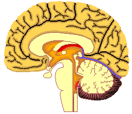
Parkinson Disease impairs an individuals muscle control. A section of the
brain called the substantia nigra contains nerve cells called nigral
cells. The nigral cells are responsible for producing a chemical called
dopamine that travels to another section of the brain called the
straitum. In the straitum, the dopamine chemical activates nerve cells
that control muscle coordination. In Parkinson Disease patients, nigral
cells die at an accelerated pace thereby reducing the amount of dopamine
available in the straitum. The destruction of nigral cells could be due
to a combination of genetics, environmental agents, and free
radicals. Patients become troubled because they cannot control their
muscle movements. This reduction of control often comes in the form of
muscle trembling, muscle inertia, impaired reflexes, and mental/physical
sluggishness. Sometimes Parkinson Disease can resemble Alzheimers
disease.
It is very difficult to simply increase the level of dopamine within the
straitum. A drug called Levodopa is structured so that it can reach the
brain and then be converted into dopamine. Only 5% of the drug reaches
the brain while the other 95% of it produces unwanted side effects such as
vomiting and nausea. Other tissues, the liver and small intestine, break
down the Levodopa before it can reach the brain. Other combinations of
drugs allow different amounts of dopamine to be produced within the
brain. Some other chemicals that are in use include agents that mock
dopamine, anticholinergics that subdues a neurotransmitter
(acetylcholine) imbalance, and amantadine a free radical production
inhibitor. As the disease progresses, it becomes unfeasible to provide
patients with enough dopamine or dopamine creating agents to suffice the
straitum. Long-term drug use creates a condition of dyskinesia and an
on-off effect.
For the patients that do not respond well to chemical therapy, surgery
has become an option. Surgical procedures can be used to destroy sections
of the brain that is infected with Parkinson Disease. It is very
difficult for doctors to pinpoint the portion of the brain that needs to
be destroyed. A deep brain stimulator device, similar to a heart
pacemaker, can be implanted into a patients brain. Instead of destroying
sections of the brain, electrical signals are sent to portions of the
brain to help control muscle movements. The implanted system requires
exterior cords from the brain to a monitoring system. This is physically
impairing and it increases a patients risk for infection. The DBSS will
locate an implementation site of an electrode for a deep brain
stimulation.
The DBSS can also assist doctors that execute surgical procedures. Two
similar procedures are used. The first, least common, is called
Thalamotomy. A small section of the thalamus that relays signals
coordinating movement is targeted for neuron extermination. A more
frequently used surgical procedure is called Pallidotomy. This operation
destroys cells in the globus pallidum, the portion of the brain that
produces uncontrolled spasmodic movements in Parkinson Disease
patients. The surgeon locates the thalamus or globus pallidum using an
MRI. A small hole is drilled into a patients skull where a tiny metal
probe is inserted deep into the brain. A burst of electricity is sent
through the probe to the targeted tissues for destruction.
Vibration stimulus applied to the Achilles tendon
Muscle vibration was used to stimulate the proprioceptors on the Achilles
tendon. The voluntary dorsiflexion movements of the ankle joint were
compared between parkinsonian and control subjects. 20 Parkinson Disease
patients that were taking Levodopa and an equal number of control subjects
were chosen. The subjects were trained to make amplitude motions of 20
degrees at 9.7 m/s. This was repeated until the subject was able to
consistently repeat the movement. Vibration at 105 hertz, 0.7-mm peak to
peak was applied to the Achilles tendon. The movement measurement was
attained after two seconds of a Go cue.
The non-vibration movements did not differ that much between
Parkinson Disease patients and control subjects. Both of the groups
suffered in movement when vibration was applied. The mean vibration to
non-vibration trials for PD and healthy subjects were, 0.86 and 0.54
respectively. The healthy subjects undershot the goals by almost 50%
while the PD group moved to around18 degrees. Charts showed that after
two seconds of vibration, the maximum proprioceptive illusion was
achieved. This shows that vibration induced errors are reduced by
Parkinson Disease.
Influence of Vibration to the Neck, Trunk and Lower Extremity Muscles
An investigation was carried out that studied the role of
proprioceptors of different skeletal muscles in postural control, in
normal subjects and patients with ULD (unilateral labyrinthine
dysfunction). The subject pool was comprised of 59 normal subjects and 12
patients with ULD. Static posturography was measured with a force
platform. The force platform was used to measure changes in the center of
gravity. The SPG data was recorded on a computer and analyzed by a signal
processor. The recording was carried out for 20 seconds.
The vibration was at 100hrtz and at amplitude of about 1mm. The
vibration was applied to the triceps, quadriceps, tibialis anterior,
biceps, and upper dorsal neck. Significant differences were found in all
muscle groups between vibration and non-vibration trials. The triceps
muscle had the largest different in SPG data between ULD patients and
healthy subjects. The Dorsal neck muscles had a small difference.
Vibratory simulation to the skeletal muscles causes instability of
standing posture in healthy patients. This is thought to occur because of
the overload of afferent data being sent from muscle spindles to the
brain. Vibration on the upper dorsal neck muscle caused an anterior body
tilt. The stimulation of the tibialis anterior created significant body
sway. The patients with ULD displayed larger sways, smaller sways, or
movement toward their body of lesions. The body movement was equal
between ULD patients and healthy subjects.
Illusional sensation of movement evoked by vibration of an immobilized arm
It takes 50 120 hertz of transcutaneous vibration of muscle spindles
primary to induce a tonic reflex. An article written by S. Rome that
investigated the perception of vibration-induced arm movement in patients
with dystonia. Dystonia is much like Parkinson Disease; both exhibit
muscle spasms and twitches. In the experiment, the arm was immobilized
because this minimizes the amount of afferent input from the joints. A
hand-held physiotherapy vibrator at 100hrtz was applied to one bicep just
above the elbow for 45 seconds. On the other free arm, patients were
instructed to copy the sensation of movement from the other arm. The
movement of the tracking was recorded by three infrared videos. This was
repeated with both arms. It was discovered that the perception of
illusional arm extension was reduced significantly and bilaterally in
patients with idiopathic focal dystonia. The angle of movement was
measured from the resting point to the maximum lift of the tracking
arm. An interesting finding was that the vibration of the biceps brachia
tendon induced muscle spasms in four patients with writers cramp.
Proprioceptive control of wrist movements in Parkinsons disease
Two groups of people, one with Parkinson Disease and the other
healthy, were compared and contrasted with respect to wrist movement. The
forearm of the subjects was rested on a horizontal support of foam
padding. It was clamped to the padding so that the only afferent input to
the brain was from wrist movement. A screen was placed between the
persons eyes and wrist so that they could not watch their movements. In a
practice session, the patients were asked to superimpose two cursors on a
monitor screen with the movement of their wrist. They were given 2.25
seconds to do so with a 5-second rest between sessions. A total of 15
practice runs was allowed.
A 100hrtz sinusoidal mechanical stimuli was applied to the tendon
of the flexor carpi radialis muscle by a small electromagnetic
vibrator. The peak-to-peak amplitude was set at 0.7mm. Each of the
subjects performed a number of vibration trials and non-vibration
trials. The Parkinson Disease patients undershot the target greater than
the healthy subjects in both vibration and non-vibration trials. The
ratio of undershooting of vibration to non-vibration trials in the healthy
group was about 0.66 while it was around 0.88 in the Parkinson Disease
group. This infers that the vibration created a larger undershooting of
the target for the Parkinson Disease subjects. The vibration reduced the
movement amplitude just over 30%, 17.25 degrees to 11.75 degrees, in the
PD group.
Circuit Construction